
Written By: Jeffrey Atlas, Health Content Writer
Medically Reviewed By: Dr. Gopal Grandhige, MD, FACS, Board-Certified Surgeon
Last Reviewed: February 10, 2026
When detecting hernias, ultrasound is the first-line imaging choice for most cases, particularly inguinal and umbilical hernias, due to its real-time visualization, safety, and lack of radiation exposure. For complex hernias or suspected complications like obstruction or strangulation, CT scans provide detailed cross-sectional imaging, while MRI offers superior soft tissue detail ideal for surgical planning. X-rays have limited direct diagnostic value but help rule out alternative conditions.
Key imaging comparison:
Ultrasound: Best for common hernias (inguinal, umbilical, femoral); no radiation; 15-30 minutes
CT Scan: Best for complex hernias and complications; uses radiation; 10-15 minutes
MRI: Best for surgical planning and soft tissue detail; no radiation; 30-60 minutes
X-ray: Limited hernia detection; minimal radiation; mainly for ruling out other conditions
Safety considerations: Ultrasound and MRI are radiation-free, making them preferred for pregnant patients and those requiring multiple scans. CT scans involve radiation exposure that should be weighed against diagnostic benefits. Your healthcare provider will select the appropriate imaging based on hernia type, complexity, and individual patient factors.

Ultrasound – Real-Time Dynamic Visualization
Medical facilities widely implement ultrasound technology as a versatile diagnostic approach for hernia identification. This technique employs high-frequency sound wave technology to generate live imaging of internal anatomical structures, enabling physicians to detect and visualize hernias with exceptional precision.
Ultrasound Advantages in Hernia Detection
Ultrasound technology successfully identifies multiple hernia classifications, including inguinal, umbilical, and femoral varieties. The dynamic visualization capability delivers critical data regarding hernia positioning, dimensions, and structural characteristics, empowering medical professionals to establish accurate diagnoses and formulate targeted treatment strategies.
Important Factors When Selecting Ultrasound
Though ultrasound demonstrates high effectiveness for specific hernia types like inguinal hernias, diagnostic precision may decrease with more complicated presentations. Complex hernia cases often require supplementary imaging approaches such as CT or MRI scanning. These advanced modalities deliver thorough assessment capabilities for intricate hernias, supplying comprehensive diagnostic data that guides effective treatment planning.
Understanding the Ultrasound Process
The ultrasound examination process remains completely painless and non-invasive. Technicians apply conductive gel to the skin covering the herniated region, ensuring optimal contact between skin surface and ultrasound equipment. A trained technician or physician manipulates a transducer device smoothly across the skin surface. This transducer releases sound waves that reflect back, generating live images displayed on monitoring screens, permitting physicians to examine and assess the hernia condition.
Safety Profile of Ultrasound Imaging
Ultrasound technology offers substantial safety advantages. Unlike alternative imaging modalities utilizing ionizing radiation, ultrasound operates exclusively with harmless sound wave frequencies. This non-invasive methodology eliminates radiation exposure entirely, establishing it as the preferred diagnostic option, particularly for expecting mothers and patients requiring multiple imaging sessions.
CT Scanning – Advanced Imaging for Complicated Hernias
CT (computed tomography) scanning represents another sophisticated imaging solution for hernia diagnosis, particularly effective for complex or internal hernia presentations. This technology merges X-ray imaging with computerized processing to produce detailed cross-sectional body imagery.
CT Scan Benefits in Hernia Assessment
CT scanning excels at detecting complicated hernias and assessing potential complications including hernia obstruction or strangulation. These scans provide extensive anatomical data that supports treatment strategy development. While ultrasound delivers real-time visualization, CT scanning offers more comprehensive hernia evaluation, including size determination, precise location mapping, and complication identification.
Key Considerations for CT Scan Selection
When evaluating imaging options for hernia diagnosis, comparing CT scanning against alternative modalities like MRI, ultrasound, and X-ray proves essential. CT scans deliver detailed cross-sectional imagery, demonstrating particular effectiveness in evaluating complex hernias and associated complications. However, radiation exposure presents a consideration.
Although radiation levels employed in CT scanning fall within generally accepted safety parameters, repeated radiation exposure potentially elevates long-term health effect risks, including cancer development possibilities.
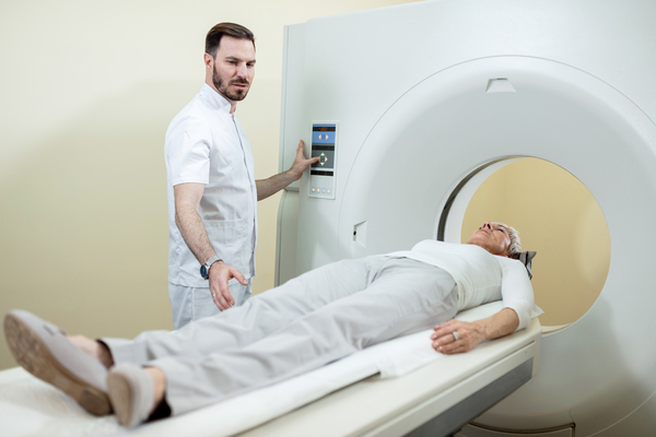
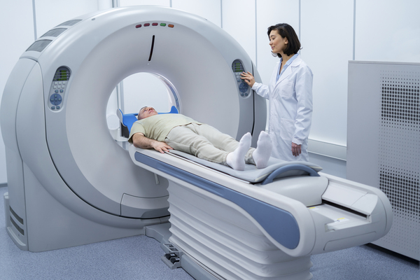
CT Scan Protocol for Hernias
CT scan procedures position patients on a movable table that advances through a circular scanning apparatus. X-ray images capture data from multiple angles, with computer systems compiling this information into detailed cross-sectional imagery. Physicians interpret these compiled images to evaluate the hernia and surrounding anatomical structures.
CT Scan Risk Factors
The principal CT scan risk involves ionizing radiation exposure. CT scanning utilizes X-ray technology to generate detailed cross-sectional body images.
Furthermore, certain individuals may experience allergic responses to contrast dye administered during CT scanning. Healthcare providers sometimes introduce contrast dye to improve visibility of specific structures or abnormalities. Contrast dye allergic reactions range from mild to severe manifestations, encompassing symptoms like skin rash, itching sensations, breathing difficulties, or tissue swelling.
Nevertheless, serious allergic reactions occur relatively infrequently.
MRI Scanning – Superior Detail and Image Clarity
Magnetic resonance imaging (MRI) stands as a highly precise imaging technique for hernia diagnosis. This modality harnesses powerful magnetic fields combined with radio wave technology to create detailed imagery of internal body structures.
MRI Scan Advantages for Hernia Diagnosis
MRI scanning provides exceptional detail and image clarity, proving particularly valuable for complex hernia cases or situations requiring precise anatomical information for surgical planning purposes.
MRI Scan Selection Considerations
MRI technology detects numerous hernia varieties, including inguinal, incisional, and hiatal classifications. This imaging method offers superior soft tissue visualization capabilities, facilitating hernia identification and characterization.
Medical professionals prefer MRI when evaluating complex hernias or associated structural abnormalities. According to Cleveland Clinic, advanced imaging techniques are essential for determining the extent of herniation and planning appropriate interventions.
MRI Scan Procedure Explained
MRI procedures position patients on a sliding table that enters a tunnel-shaped scanning device. The apparatus generates powerful magnetic fields and radio waves that create detailed internal structure imagery. Physicians analyze these images to evaluate the hernia and related anatomical structures.
MRI Scan Risk Assessment
MRI scanning maintains a generally safe profile without ionizing radiation exposure. However, patients with specific metallic implants or medical devices may not qualify as suitable MRI candidates due to the powerful magnetic field environment.
Certain patients may experience claustrophobia or anxiety reactions within the MRI machine’s enclosed environment. Healthcare teams provide support and management strategies to address these concerns throughout the scanning process.
Contrast agents, particularly gadolinium-based compounds, may be utilized in select cases to enhance specific structure or abnormality visibility. While adverse reactions to these contrast materials remain rare, minimal risks of allergic reactions or alternative side effects exist. Patients should communicate any known allergies or previous contrast agent reactions to healthcare providers before undergoing MRI scanning.

X-ray Imaging – Restricted Role in Hernia Diagnosis
X-ray imaging, though not typically serving as the primary hernia diagnostic tool, can occasionally reveal indirect indicators of certain hernia types.
X-ray Selection Considerations
X-ray technology proves more suitable for identifying complications or excluding alternative conditions that may present similarly to hernias, such as bowel obstruction or pneumoperitoneum (abdominal cavity air presence).
X-rays involve minimal radiation exposure levels and maintain a safe profile. However, their direct hernia visualization application remains limited compared to ultrasound, CT scanning, or MRI technology. Situations requiring more detailed evaluation necessitate alternative imaging approaches.
X-ray Procedure for Hernia Assessment
X-rays provide limited direct diagnostic application for hernias but offer utility in specific circumstances. X-ray procedures involve radiographer positioning of patients, followed by machine emission of minimal radiation amounts to capture internal structure images. Healthcare professionals interpret these X-ray images to assess for indirect hernia indicators or related complications.
X-ray Risk Evaluation
X-ray imaging involves minimal radiation exposure with generally negligible associated risks. However, maintaining awareness of radiation exposure remains important, especially when repeated X-rays or multiple imaging procedures become necessary. Healthcare providers ensure X-ray benefits outweigh potential risks. Pregnant patients should specifically inform healthcare providers regarding pregnancy status, as special precautions may prove necessary to minimize fetal radiation exposure.
Conclusion
Selecting the appropriate imaging method for hernia detection requires careful consideration of the hernia type, complexity, and individual patient factors. Ultrasound remains the preferred first-line diagnostic tool due to its safety profile, real-time visualization capabilities, and effectiveness in detecting common hernias like inguinal and umbilical varieties. For more complex cases or when complications are suspected, CT scans provide comprehensive cross-sectional imaging that helps identify obstruction or strangulation, though radiation exposure must be weighed against diagnostic benefits.
MRI offers superior soft tissue detail without radiation, making it ideal for surgical planning and evaluating intricate hernia presentations, particularly when anatomical precision is paramount. Hiatal hernias, in particular, often benefit from advanced imaging techniques to assess their relationship with surrounding digestive structures. Research from Johns Hopkins Medicine emphasizes the importance of selecting the right diagnostic approach for optimal patient outcomes.
While X-rays play a limited direct role in hernia diagnosis, they remain useful for ruling out alternative conditions or identifying complications. For patients experiencing symptoms like GERD or heartburn related to abdominal hernias, proper imaging is crucial for determining whether a hernia is contributing to digestive issues. The National Institute of Diabetes and Digestive and Kidney Diseases provides comprehensive information on how hernias affect digestive function.
Understanding your diagnostic options is essential when dealing with conditions that may coexist with hernias, such as achalasia, gastroparesis, or silent reflux. According to Harvard Health, accurate imaging enables physicians to differentiate between various gastrointestinal conditions that may present similar symptoms.
Once a hernia is properly diagnosed through imaging, various surgical procedures may be recommended depending on the severity and type. Modern treatment options include LINX procedures and TIF with EsophyX for certain types of hernias affecting the esophageal region. For patients also concerned about weight management, which can impact hernia development and treatment outcomes, minimally invasive weight loss options may be discussed alongside hernia repair strategies. WebMD offers additional information about how imaging results guide treatment selection.
For patients seeking comprehensive care, consulting with experienced hernia specialists ensures proper interpretation of imaging results and development of personalized treatment plans. The NHS recommends working closely with healthcare teams to understand imaging findings and their implications for treatment.
Ultimately, healthcare providers determine the most appropriate imaging modality based on clinical presentation, ensuring accurate diagnosis and optimal treatment planning for each patient’s unique situation. Additional resources from Healthline, MedlinePlus, and NCBI provide further evidence-based information about hernia diagnosis and management. To learn more about diagnostic options or schedule a consultation, patients can explore comprehensive resources available through specialized digestive health centers.
FAQs
Which imaging test is best for detecting inguinal hernias?
Ultrasound is typically the most effective and preferred imaging method for detecting inguinal hernias due to its real-time visualization capabilities and high accuracy. It’s also safe, non-invasive, and doesn’t involve radiation exposure.
Do all hernia imaging tests involve radiation exposure?
No, ultrasound and MRI do not use ionizing radiation and are completely radiation-free. CT scans and X-rays do involve radiation exposure, though the levels are generally considered safe for diagnostic purposes.
How long does each imaging procedure typically take?
Ultrasound examinations usually take 15-30 minutes, while CT scans are typically completed in 10-15 minutes. MRI procedures generally require 30-60 minutes due to the detailed imaging process involved.
Can pregnant women safely undergo hernia imaging?
Ultrasound is the safest option for pregnant women as it uses only sound waves without radiation exposure. MRI is generally safe after the first trimester, while CT scans and X-rays should be avoided unless absolutely necessary.
Will I need contrast dye for my hernia imaging test?
Contrast dye is rarely needed for ultrasound examinations but may be used in some CT and MRI scans to enhance visualization. Your healthcare provider will inform you in advance if contrast is necessary and screen for potential allergies.


An endoscopy cannot tell you if you have reflux. It can only tell you if you have complications of GERD.
If you are unhappy with your reflux symptoms, come in and we can discuss testing and treatments that can accurately diagnose your problem.
#reflux #gerd #hiatalhernia #gastroparesis #linx

CALL US AT 813-922-2920
www.tampareflux.com
If you have a hiatal hernia and fit one of these categories, you should know your options.
Dr. Grandhige is an expert in his field and performs 200 of these surgeries a year. He is the only surgeon in the Tampa Bay Area who offers all surgical options - LINX, Fundoplications, TIF and will be one of 20 surgeons in America introducing the latest procedure RefluxStop in 2026.
We accept most insurances but will verify yours before you come in. These procedures are considered medically necessary and covered by your insurance. You can expect to pay your in-network deductibles and nothing else.
#hiatalhernia #reflux #GERD #LINX #refluxstop
What causes reflux ?
1. Weak lower esophageal sphincter
2. Hiatal hernia
3. Flattening of the Angle of His
4. Poor esophageal motility
5. Gastroparesis (slow stomach)
NOT increased acid production
Don’t let GERD get in the way of living your life. Request your appointment with us today on the link below.
.
.
.
.
https://tampareflux.com/contact-us/
Anyone can be victim to GERD and though weight loss can help reduce GERD symptoms. Many athletes with high impact workouts may continue to have these symptoms. This may be a symptom of a hiatal hernia or other issue. We are more then happy to assist you in finding your solution, just click the link below.
.
.
.
https://tampareflux.com/contact-us/
##healthylifestyle #workout #athletereflux #PPIs #heartburn #LINX #fundoplication #TIF #GERD#tampaheartburn #linx #TIF #fundoplication #tampabayreflux #GERD #acidreflux #acidrefluxsurgery #stopreflux
#nonsurgicalweightloss #ESG #gastricballoon #weightlossjourney #vsg #vsgjourney #spatz3 #orbera #orberaballoon #grandhige #DrG
#tampabayrefluxinstitute #guthealth #roboticsurgery
Heartburn may seem like an annoyance. But if you find yourself having symptoms on a daily basis, it may be time to to talk to Dr. Grandhige as it could be a symptom of something worse.
.
.
.
#chronicheartburn #gerdsymptoms #heartburnrelief #reflux #PPIs #heartburn #LINX #fundoplication #TIF #GERD#tampaheartburn #linx #TIF #fundoplication #tampabayreflux #GERD #acidreflux #acidrefluxsurgery #stopreflux
#nonsurgicalweightloss #ESG #gastricballoon #weightlossjourney #vsg #vsgjourney #spatz3 #orbera #orberaballoon #grandhige #DrG
#tampabayrefluxinstitute #guthealth #roboticsurgery
If you are tired of avoiding your favorite foods or taking daily medications, we can help.
We are the Tampa experts in reflux ! With years of experience and thousands of patients treated successfully, we offer all FDA approved anti-reflux procedures.
Call 813-922-2920 to schedule your appointment
All major insurances accepted.
Not all patients need surgical intervention. Many patients are living a heartburn free life with their PPIs. However 40% of patients taking PPIs are not getting the relief they need. If you are one of those, you have options! Come in and find out more.
.
.
.
.
#letushelpyou #medsnotworking #reflux #PPIs #heartburn #LINX #fundoplication #TIF #GERD#tampaheartburn #linx #TIF #fundoplication #tampabayreflux #GERD #acidreflux #acidrefluxsurgery #stopreflux
#nonsurgicalweightloss #ESG #gastricballoon #weightlossjourney #vsg #vsgjourney #spatz3 #orbera #orberaballoon #grandhige #DrG
#tampabayrefluxinstitute #guthealth #roboticsurgery
#heartburn #stopreflux #hiatalherniarepair #severeheartburn #reflux #tampabayreflux #acidrefluxsurgery #tampaheartburn #GERD #PPIs #achalasia #LINX #TIF #tampareflux #fundoplication #stomach #digestivehealth #ESG #obesity #overweight #weightlossjourney #gastricballoon