
Written By: Dr. Ahmad Saad, Health Content Writer
Medically Reviewed By: Dr. Gopal Grandhige, MD, FACS, Board-Certified Surgeon
Last Reviewed: January 17, 2026
Gastroparesis is diagnosed through a combination of clinical evaluation and specialized testing that confirms delayed gastric emptying without mechanical obstruction. The diagnostic process typically requires:
Primary Diagnostic Criteria:
- Symptoms consistent with gastroparesis (nausea, vomiting, early satiety, abdominal discomfort)
- Absence of physical blockage in the stomach
- Documented delayed gastric transit through objective testing
The 5 Key Diagnostic Tests:
- Gastric Emptying Scintigraphy (GES), Gold standard test using radioactive tracers to measure stomach emptying speed over 2-4 hours
- Upper GI Endoscopy, Visual examination to rule out obstructions and identify retained food
- Laboratory Tests, Blood and urine analysis to detect diabetes, electrolyte imbalances, and exclude other conditions
- Wireless Motility Capsule (SmartPill), Ingestible device measuring pH, pressure, and transit times throughout the digestive system
- Electrogastrography (EGG), Non-invasive test recording stomach electrical activity patterns
Diagnosis Timeline: The complete diagnostic process typically takes several weeks to months, involving sequential testing procedures tailored to individual symptoms.
Most Reliable Test: Gastric emptying scintigraphy achieves the highest diagnostic accuracy, with delayed emptying defined as ≥30% meal retention at 1 hour, ≥60% at 2 hours, or ≥10% at 4 hours.

Gastroparesis Explained: A Chronic Digestive Condition
Gastroparesis represents a persistent disorder compromising the stomach’s capacity to transfer contents into the small bowel. Grasping its nature, manifestations, and underlying causes proves vital for patients.
Defining Gastroparesis
This motility disorder means the stomach requires excessive time to evacuate its contents, occurring without physical obstruction present.
Gastric motility depends on coordinated muscle and nerve function. Gastroparesis disrupts this coordination, resulting in prolonged gastric transit times.
Primary Symptoms and Red Flags
Gastroparesis manifestations differ among individuals but commonly feature:
- Persistent nausea with episodes of vomiting
- Post-meal fullness or abdominal bloating
- Upper stomach discomfort or pain sensations
- Rapid satiety during eating
- Unintended weight reduction and nutritional deficiencies
These manifestations significantly compromise daily functioning and quality of life, warranting prompt medical consultation when persistent.
Underlying Causes and Contributing Factors
Multiple factors can trigger gastroparesis development:
- Diabetic complications
- Surgical procedure aftereffects
- Nerve-related disorders
- Specific pharmaceutical agents
- Unknown origins (idiopathic presentation)
Identifying the root cause proves essential for condition management. Contributing factors encompass longstanding diabetic disease, specific neurological conditions, and prior abdominal surgical procedures. According to the National Institute of Diabetes and Digestive and Kidney Diseases, diabetes is one of the most common known causes of gastroparesis.
The Critical Role of Precise Diagnosis
Obtaining accurate gastroparesis diagnosis carries immense significance. This complex condition demands exact identification for appropriate therapeutic intervention.
The Value of Correct Diagnosis
Precise diagnostic findings enable physicians to distinguish gastroparesis from other disorders presenting similar symptoms. Accurate identification ensures patients receive targeted treatments, substantially enhancing quality of life. Misdiagnosis leads to ineffective therapeutic approaches that fail to address the actual problem.
Key reasons accurate diagnosis matters:
- Enables appropriate therapeutic strategies
- Minimizes complication risks
- Enhances patient treatment outcomes
- Facilitates patient understanding of their condition
Disorders Resembling Gastroparesis
Several conditions can mimic gastroparesis presentation, complicating diagnostic efforts:
- Functional dyspepsia syndrome
- Cyclic vomiting disorder
- Acid reflux disease (GERD)
- Peptic ulcer conditions
Medical professionals must recognize these similar conditions, employing combined clinical evaluation and diagnostic testing to establish correct diagnoses. Conditions like hiatal hernias may also produce overlapping symptoms requiring careful differentiation.
The Diagnostic Process
Gastroparesis diagnosis involves multiple sequential steps:
- Initial comprehensive clinical evaluation
- Laboratory analysis
- Diagnostic imaging procedures
- Specialized tests including gastric emptying scintigraphy
Patients typically undergo numerous evaluations to confirm gastroparesis presence. Persistence and patience prove necessary throughout this process. Accurate diagnosis establishes the foundation for effective condition management.
Recognizing the importance of diagnostic precision helps both healthcare providers and patients successfully navigate this evaluation journey.
Initial Medical Evaluation
Gastroparesis diagnosis begins with thorough initial clinical evaluation, encompassing several critical components essential for understanding patient status and directing subsequent testing.
Medical Background Review
Comprehensive medical history collection represents the initial step. Providers gather information regarding symptom presentation, duration, and severity levels. Additional inquiry covers dietary patterns, current medications, and previous health conditions. Understanding medical background helps identify potential gastroparesis causes and exclude alternative diagnoses.
Surgical history receives particular attention, as certain operations may precipitate gastroparesis development. Providers investigate underlying conditions including diabetes or neurological disorders, which show strong gastroparesis associations. Patients who have undergone fundoplication procedures should inform their physicians, as post-surgical gastroparesis can sometimes occur.
Physical Assessment
Complete physical examination proves equally essential. Clinicians evaluate for malnutrition signs, dehydration indicators, or other complications. Examination includes checking for abdominal tenderness or additional abnormalities potentially causing symptoms.
Physical findings provide crucial information that, combined with medical history, creates comprehensive understanding of patient condition and helps pinpoint underlying causes.
Symptom Documentation and Journals
Symptom tracking through patient diaries represents valuable diagnostic tools. Providers may request patients maintain detailed journals documenting food intake, symptom patterns, and other relevant observations. This practice reveals patterns and identifies potential symptom triggers.
Patient journals offer invaluable insights, enabling tailored diagnostic testing and treatment planning. Through careful initial assessment analysis, clinicians develop more accurate diagnoses and effective gastroparesis management strategies.
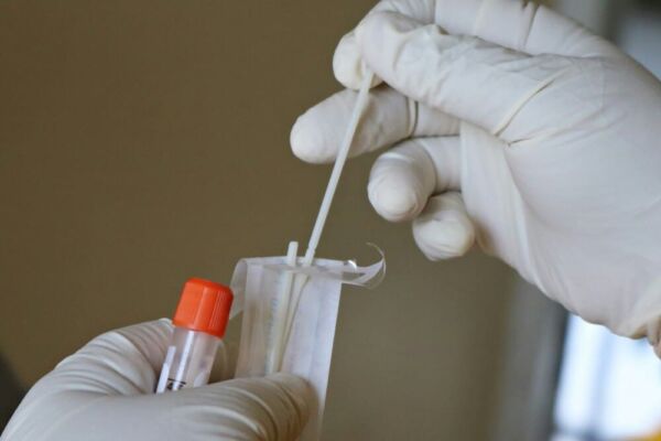
Laboratory Analysis for Gastroparesis
Laboratory testing plays a fundamental role in gastroparesis diagnosis, helping physicians exclude other conditions with comparable symptoms. These analyses prove vital for identifying symptom causes and confirming gastroparesis presence.
Blood Analysis
Blood testing constitutes a major diagnostic component for gastroparesis. These tests evaluate overall health status and screen for underlying conditions. Common analyses include:
- Complete Blood Count (CBC): Identifies infection or inflammation indicators
- Glucose Level Tests: Evaluates blood sugar concentrations, since diabetes commonly causes gastroparesis
- Electrolyte Panels: Checks for imbalances in critical minerals including potassium and sodium
Urine Testing
Urinalysis represents another crucial gastroparesis diagnostic test, analyzing urine samples to detect underlying conditions. Testing can identify:
- Ketoacidosis: A diabetic complication producing gastroparesis-like symptoms
- Urinary Tract Infections: Which may generate symptoms resembling gastroparesis
- Additional Abnormalities: Including blood or protein presence in urine
Excluding Alternative Diagnoses
Laboratory testing proves vital for eliminating other conditions potentially causing similar manifestations. Through these analyses, physicians can:
- Detect Underlying Conditions: Including diabetes, thyroid dysfunction, or kidney disorders
- Rule Out Alternative Causes: Of symptoms, such as ulcerative conditions or gastritis
- Confirm Accurate Diagnosis: Of gastroparesis based on laboratory findings and additional diagnostic procedures
By integrating laboratory results with other diagnostic methods, physicians establish accurate diagnoses and develop effective gastroparesis treatment protocols. The Cleveland Clinic emphasizes the importance of comprehensive testing in achieving proper diagnosis.
Gastroparesis Diagnosis: The Premier Testing Method
Achieving precise gastroparesis diagnosis proves essential for successful management. The gold-standard diagnostic test remains Gastric Emptying Scintigraphy (GES), which measures gastric transit speed, revealing stomach function status.
Gastric Emptying Scintigraphy (GES)
Gastric Emptying Scintigraphy utilizes nuclear medicine imaging to assess stomach-to-small intestine food transfer rates. Patients consume a meal containing minimal radioactive material, typically technetium-99m incorporated into scrambled eggs or pancakes.
Preparation and Testing Protocol
Pre-test preparation requires overnight fasting. Following this, patients consume the radioactive meal. A specialized camera captures abdominal images at designated intervals, typically 0, 1, 2, and occasionally 4 hours post-consumption. This tracks gastric emptying velocity.
Understanding GES Findings
Test results demonstrate radioactive material gastric clearance rates. Prolonged retention may indicate gastroparesis presence. Results show meal retention percentages in the stomach at various timepoints, supporting diagnostic conclusions.
| Time (Hours) | Normal Gastric Transit (%) | Delayed Gastric Transit (%) |
| 1 | < 30% | ≥ 30% |
| 2 | < 60% | ≥ 60% |
| 4 | < 10% | ≥ 10% |
Benefits and Drawbacks
GES represents the premier test due to superior accuracy in gastroparesis identification. However, testing involves minimal radiation exposure and limited facility availability. Despite these limitations, GES remains an indispensable gastroparesis diagnostic tool.
In summary, Gastric Emptying Scintigraphy provides unparalleled insights into gastric function. Understanding this testing methodology enables physicians to establish better diagnoses and create effective treatment strategies for gastroparesis patients. The Mayo Clinic provides additional resources on this diagnostic procedure.
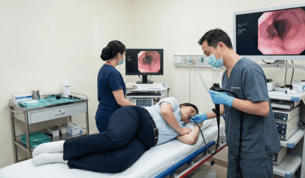
Upper Digestive Endoscopy
Upper GI endoscopy serves a crucial role in gastroparesis diagnosis, allowing physicians to visualize upper digestive tract structures. This proves essential for detecting obstructions and evaluating upper digestive system health.
Objective and Pre-Procedure Requirements
The primary purpose of upper GI endoscopy in gastroparesis evaluation involves examining the esophagus, stomach, and proximal small intestine. This identifies problems including blockages or inflammatory conditions potentially causing gastroparesis-like symptoms.
Pre-procedure preparation typically requires several hours of fasting. Procedures occur under sedation for patient comfort. Physicians may request temporary discontinuation of certain medications that could interfere with test accuracy.
Endoscopic Examination Process
A flexible tube equipped with camera and lighting (endoscope) enters through the mouth, advancing through the esophagus into stomach and duodenum. The endoscope transmits images to a monitor, enabling physicians to examine upper GI tract lining for abnormalities.
Complete procedures typically require 30 minutes to one hour. Some patients experience mild discomfort or temporary throat soreness afterward, though these symptoms generally resolve quickly.
Gastroparesis Appearance During Endoscopy
During gastroparesis endoscopy, physicians seek signs of delayed gastric transit or associated conditions. Gastroparesis itself may lack distinct endoscopic findings. However, testing can reveal retained food in the stomach, a significant gastroparesis indicator.
Tissue Sampling and Additional Testing
Physicians may obtain biopsies during endoscopy, small stomach lining tissue samples examined microscopically for inflammation, infection, or other abnormalities.
Endoscopy and biopsy findings, combined with additional diagnostic tests, enable physicians to accurately diagnose gastroparesis and formulate evidence-based treatment plans. These procedures are also valuable for evaluating related conditions like achalasia and silent reflux (LPR).
| Diagnostic Element | Description | Gastroparesis Relevance |
| Endoscopy Purpose | Upper GI tract examination | Excludes mechanical blockages and alternative conditions |
| Preparation | Fasting, medication adjustments | Ensures result accuracy and patient safety |
| Procedure | Endoscope insertion, visualization | Identifies abnormalities, food retention |
| Biopsy | Tissue collection for microscopic analysis | Excludes conditions like gastritis |
Additional Diagnostic Approaches
Beyond conventional testing methods, alternative diagnostic approaches for gastroparesis exist. While gastric emptying scintigraphy remains the gold standard, supplementary tests provide valuable complementary information, enhancing understanding and enabling optimized treatment planning.
Gastric Transit Breath Analysis
Gastric emptying breath tests offer simple, non-invasive assessment of stomach emptying rates. Patients consume meals containing special substances, with subsequent breath analysis detecting these markers.
Benefits: Simple procedure, radiation-free, office-based testing capability
Wireless Motility Monitoring (SmartPill)
The wireless motility capsule (SmartPill) represents an ingestible miniature device monitoring pH, pressure, and temperature throughout the digestive system. This technology reveals transit times through stomach, small intestine, and colon.
Antroduodenal Pressure Measurement
Antroduodenal manometry evaluates stomach and duodenum motility function. A thin catheter inserted through nose or mouth into these regions contains sensors measuring muscle contraction strength.
This testing excels at identifying motility dysfunction, demonstrating gastric muscle coordination effectiveness. Research published in the American Journal of Gastroenterology has validated the effectiveness of these advanced diagnostic techniques.
Stomach Electrical Activity Recording (EGG)
Electrogastrography (EGG) examines stomach electrical activity patterns. Abdominal surface electrodes capture gastric electrical signals.
EGG identifies abnormal stomach rhythms and additional motility disturbances potentially causing gastroparesis symptoms. For comprehensive guidance on managing gastroparesis symptoms, the NCBI Bookshelf provides detailed clinical information.
| Diagnostic Method | Description | Primary Advantages |
| Gastric Transit Breath Test | Measures emptying rates through breath analysis | Non-invasive, radiation-free |
| Wireless Motility Capsule (SmartPill) | Records pH, pressure, temperature throughout GI tract | Comprehensive motility data, single-use device |
| Antroduodenal Manometry | Measures stomach and duodenum motility | Detailed contraction information |
| Electrogastrography (EGG) | Records stomach electrical activity | Non-invasive, detects rhythm abnormalities |
These testing methods expand diagnostic options for gastroparesis evaluation and treatment. Understanding each test’s capabilities enables physicians to select optimal approaches for individual patients.
Diagnostic Imaging for Gastroparesis
Imaging studies play vital roles in gastroparesis diagnosis, helping eliminate other conditions presenting similar symptoms. These tests deliver important information supporting diagnosis and identifying potential complications.
Ultrasonography
Ultrasound represents non-invasive testing using sound waves to generate internal organ images. This can identify gallstones, liver pathology, or other issues potentially causing symptoms in gastroparesis patients.
Benefits: Non-invasive approach, zero radiation exposure, cost-effective
Drawbacks: Operator skill-dependent, reduced effectiveness in obese patients or with intestinal gas interference
Computed Tomography Scanning
CT scanning employs X-ray technology and computer processing to produce detailed body images. This helps detect bowel obstructions, tumor masses, or other structural abnormalities in gastroparesis patients.
Benefits: Provides detailed imaging, identifies diverse abnormalities
Drawbacks: Involves radiation exposure, may require contrast administration
Magnetic Resonance Imaging
MRI utilizes powerful magnetic fields and radio waves generating detailed internal organ images. This evaluates stomach and small intestine for structural abnormalities or inflammatory conditions.
Benefits: Radiation-free imaging, produces detailed soft tissue visualization
Drawbacks: Unsuitable for patients with certain metallic implants, expensive testing
Barium Contrast X-rays
Barium X-rays involve consuming barium solution to visualize upper digestive structures. This testing identifies structural problems including narrowing or obstructions in stomach or intestinal regions.
Benefits: Provides functional digestive tract information, identifies structural issues
Drawbacks: Involves radiation exposure, potentially unsuitable for patients with specific allergies or sensitivities
Summary of imaging methods used in gastroparesis diagnosis:
| Imaging Method | Benefits | Drawbacks |
| Ultrasonography | Non-invasive, radiation-free | Operator-dependent, obesity or gas limitations |
| CT Scanning | Detailed imaging, detects various abnormalities | Radiation exposure, may require contrast |
| MRI | Radiation-free, detailed soft tissue visualization | Metal implant restrictions, expensive |
| Barium X-rays | Functional information, identifies structural issues | Radiation exposure, potential allergy concerns |
Conclusion
Accurate gastroparesis diagnosis requires a comprehensive, multi-faceted approach combining clinical evaluation, laboratory testing, and specialized diagnostic procedures. Gastric emptying scintigraphy remains the gold-standard test, providing definitive evidence of delayed gastric transit that characterizes this challenging digestive disorder. However, successful diagnosis often depends on integrating multiple testing methods, from upper GI endoscopy to wireless motility monitoring, tailored to each patient’s unique presentation and symptoms.
The diagnostic journey may seem lengthy and complex, but persistence through this process proves essential for achieving accurate identification and effective treatment outcomes. By working closely with experienced gastroenterologists and utilizing state-of-the-art diagnostic tools, patients can obtain the precise diagnosis needed to develop targeted management strategies. Early and accurate diagnosis not only improves quality of life but also helps prevent potential complications, enabling patients to regain control over their digestive health and overall wellbeing.
For patients exploring treatment options for gastroparesis, procedures like the LINX Reflux Management System and TIF EsophyX may be discussed alongside traditional management approaches. Additionally, those dealing with weight-related digestive issues might benefit from learning about incisionless weight loss procedures.
To learn more about gastroparesis diagnosis and treatment options, visit our comprehensive blog resources or contact our team for personalized guidance. For those seeking additional medical information, MedlinePlus offers detailed health encyclopedia entries on gastroparesis. Patient advocacy organizations like G-PACT and the resources at About Gastroparesis provide valuable support networks and educational materials. Peer-reviewed research is available through PMC for those interested in the latest scientific findings. Whether you’re newly diagnosed or seeking better management of your condition, Tampa Reflux Center is committed to providing compassionate, expert care.
FAQs
What is the most accurate test for diagnosing gastroparesis?
Gastric emptying scintigraphy (GES) is the gold-standard test for gastroparesis, using nuclear medicine imaging to measure how quickly food leaves your stomach. This test provides the most reliable evidence of delayed gastric transit, which is the hallmark of gastroparesis.
How long does it take to diagnose gastroparesis?
The diagnostic process typically takes several weeks to months, involving multiple appointments and sequential testing procedures. Your healthcare team will conduct initial evaluations, laboratory tests, and specialized diagnostic procedures to confirm gastroparesis and rule out other conditions.
Can gastroparesis be detected through regular blood tests?
Blood tests alone cannot diagnose gastroparesis, but they help identify underlying causes like diabetes and rule out other conditions with similar symptoms. Definitive gastroparesis diagnosis requires specialized tests like gastric emptying scintigraphy that measure stomach function directly.
Is endoscopy necessary for gastroparesis diagnosis?
Upper GI endoscopy is often performed to exclude mechanical obstructions and other conditions that mimic gastroparesis symptoms, though it cannot diagnose gastroparesis itself. The procedure helps physicians visualize your digestive tract and may reveal retained food in the stomach, supporting the diagnosis.
Are gastroparesis diagnostic tests painful or risky?
Most gastroparesis tests are non-invasive or minimally invasive with low risk, though some may cause mild discomfort. Gastric emptying scintigraphy involves minimal radiation exposure, while endoscopy is performed under sedation to ensure patient comfort throughout the procedure.


An endoscopy cannot tell you if you have reflux. It can only tell you if you have complications of GERD.
If you are unhappy with your reflux symptoms, come in and we can discuss testing and treatments that can accurately diagnose your problem.
#reflux #gerd #hiatalhernia #gastroparesis #linx

CALL US AT 813-922-2920
www.tampareflux.com
If you have a hiatal hernia and fit one of these categories, you should know your options.
Dr. Grandhige is an expert in his field and performs 200 of these surgeries a year. He is the only surgeon in the Tampa Bay Area who offers all surgical options - LINX, Fundoplications, TIF and will be one of 20 surgeons in America introducing the latest procedure RefluxStop in 2026.
We accept most insurances but will verify yours before you come in. These procedures are considered medically necessary and covered by your insurance. You can expect to pay your in-network deductibles and nothing else.
#hiatalhernia #reflux #GERD #LINX #refluxstop
What causes reflux ?
1. Weak lower esophageal sphincter
2. Hiatal hernia
3. Flattening of the Angle of His
4. Poor esophageal motility
5. Gastroparesis (slow stomach)
NOT increased acid production
Don’t let GERD get in the way of living your life. Request your appointment with us today on the link below.
.
.
.
.
https://tampareflux.com/contact-us/
Anyone can be victim to GERD and though weight loss can help reduce GERD symptoms. Many athletes with high impact workouts may continue to have these symptoms. This may be a symptom of a hiatal hernia or other issue. We are more then happy to assist you in finding your solution, just click the link below.
.
.
.
https://tampareflux.com/contact-us/
##healthylifestyle #workout #athletereflux #PPIs #heartburn #LINX #fundoplication #TIF #GERD#tampaheartburn #linx #TIF #fundoplication #tampabayreflux #GERD #acidreflux #acidrefluxsurgery #stopreflux
#nonsurgicalweightloss #ESG #gastricballoon #weightlossjourney #vsg #vsgjourney #spatz3 #orbera #orberaballoon #grandhige #DrG
#tampabayrefluxinstitute #guthealth #roboticsurgery
Heartburn may seem like an annoyance. But if you find yourself having symptoms on a daily basis, it may be time to to talk to Dr. Grandhige as it could be a symptom of something worse.
.
.
.
#chronicheartburn #gerdsymptoms #heartburnrelief #reflux #PPIs #heartburn #LINX #fundoplication #TIF #GERD#tampaheartburn #linx #TIF #fundoplication #tampabayreflux #GERD #acidreflux #acidrefluxsurgery #stopreflux
#nonsurgicalweightloss #ESG #gastricballoon #weightlossjourney #vsg #vsgjourney #spatz3 #orbera #orberaballoon #grandhige #DrG
#tampabayrefluxinstitute #guthealth #roboticsurgery
If you are tired of avoiding your favorite foods or taking daily medications, we can help.
We are the Tampa experts in reflux ! With years of experience and thousands of patients treated successfully, we offer all FDA approved anti-reflux procedures.
Call 813-922-2920 to schedule your appointment
All major insurances accepted.
Not all patients need surgical intervention. Many patients are living a heartburn free life with their PPIs. However 40% of patients taking PPIs are not getting the relief they need. If you are one of those, you have options! Come in and find out more.
.
.
.
.
#letushelpyou #medsnotworking #reflux #PPIs #heartburn #LINX #fundoplication #TIF #GERD#tampaheartburn #linx #TIF #fundoplication #tampabayreflux #GERD #acidreflux #acidrefluxsurgery #stopreflux
#nonsurgicalweightloss #ESG #gastricballoon #weightlossjourney #vsg #vsgjourney #spatz3 #orbera #orberaballoon #grandhige #DrG
#tampabayrefluxinstitute #guthealth #roboticsurgery
#heartburn #stopreflux #hiatalherniarepair #severeheartburn #reflux #tampabayreflux #acidrefluxsurgery #tampaheartburn #GERD #PPIs #achalasia #LINX #TIF #tampareflux #fundoplication #stomach #digestivehealth #ESG #obesity #overweight #weightlossjourney #gastricballoon